Study could lead to new therapies to help improve sight following trauma or stroke
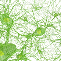 University of Texas neuroscientists having been looking at how nerve cells in the visual cortex of the brain handle and adapt to images as they change.
Researchers evaluated the results of stimulating the visual cortex upon optic neurons whose electrical activity was measured at the same time in lab animals. With the animal viewing movies they monitored the behavior of visual cortex neurons as the images changed.
Results showed that short exposure or adaptation to a fixed visual stimulus caused changes in how much individual neurons cooperated with each other and in so doing improved the efficiency of the cells to encode information for interpretation by the brain.
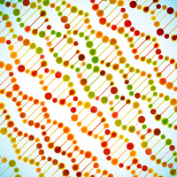
The authors of the study wrote that how we see our environment depends upon the ability of the neural networks of our brain and body to adapt very quickly to changes in what we perceive. Scientists are increasingly realizing that how our neural networks are structured and how they communicate is itself an adaptive process – our nerve cells change how they respond appropriately depending on what is in our sensory environment – converting ” electrical impulses in the brain into thoughts, memories and decisions”.
Source: “Populations Of Brain Cells Adapt To Changing Images,” Dragoi, et al., Nature 452, 220-224 (13 March 2008).
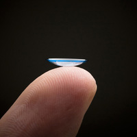 An increase in the use of contact lenses may be making ulcers of the cornea twice as common. A study of over a million Californians showed that people who wore contact lenses were 9 times more likely to suffer from corneal ulcers. Many people do not follow basic contact lens safety principles.
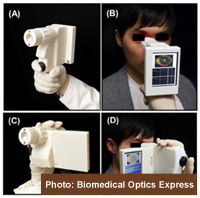 Currently, most eye disease detection equipment is only available in an optometrist’s office. A new handheld optical device could allow general practitioners to quickly screen all patients for eye disease such as glaucoma and macular degeneration. These visually devastating diseases are most easily treated or controlled in their early stages. The new high-speed prototypes were developed at Massachusetts Institute of Technology.
 Devastating eye diseases such as age-related macular degeneration (AMD) and diabetic retinopathy may be detectable sooner with a new camera being developed by ophthalmologist Jean-Daniel Arbour and Photon Etc. in Canada. The camera is designed to detect changes at the metabolic level, even before anatomical changes are visible. It uses hyperspectral photography, which utilizes all wavelengths to detect more details.
 The medications tested were proton pump inhibitors called Prilosec (Omeprazole), Nexium (Esomeprazole Magnesium) and Prevacid (Lansoprazole).