Iritis, the most common form of uveitis, is more prevalent than most people realize. What are the symptoms of this eye condition? What can you do to prevent and manage all types of uveitis? When does uveitis signal an underlying condition?
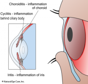
Uveitis is an inflammation of the uvea. The uvea is made up of the iris, the colored part of the eye; the ciliary body, which makes the fluid that fills the eye and flexes the eye lens; and the choroid, the layer beneath the retina.
 Types of Uveitis
-
- Iritis or anterior (front) uveitis. Anterior uveitis can involve the iris, ciliary body, cornea, and sclera. It is the most common type of uveitis and accounts for about 50–60% of all uveitis cases in special care clinics.[1. Albert, D.M., Jakobiec, F.A., editors. (2000). Principles and Practice of Ophthalmology. 2nd ed. Philadelphia, PA: WB Saunders Co.] Sixty-five percent of cases are related to another health condition. The remaining 35% are idiopathic (no discernible relationship to another health problem).
- Cyclitis or intermediate uveitis. Intermediate uveitis is the least common type of uveitis, involving the area between the ciliary body and the back of the eyeball. It has been found to account for 3–17% of uveitis around the world.[2. Ibid. Albert. (2000).]
- Choroiditis or posterior (back) uveitis. This category accounts for only 10–40% of uveitis cases. However, more visual loss results in these cases than from other uveitis forms. Such vision loss may be due to cystoid macular edema, retinal detachment, subretinal fibrosis, or optic nerve damage.[3. Jabs, D.A., Akpek, E.K. (2005). Immunosuppression for posterior uveitis Retina, Jan; 25(1):1-18.3.] Up to 50% of patients with posterior uveitis have an associated systemic disease.
 Lutein, zeaxanthin, and meso-zeaxanthin are potent antioxidants found in green leafy vegetables, eggs, corn and even chocolate. They have a profound effect on preserving healthy vision, brain function, the immune and cardiovascular systems.
 The cornea is in front of the lens. It is a clear layer, seeming to lack substance. However, it is comprised of multiple layers and groups of cells and proteins which are highly organized.
 Hypertension increases sharply with advancing age; hence older persons are those most affected by its negative consequences.
 Approximately eight per cent of men and one-half of one per cent of women in the U.S. have a problem with their color perception.
 Microcurrent Stimulation treatment protocols are designed to help people with retinal disease. MCS works by supporting nourishment and healing to the back of the eyes as well as possibly some cell regeneration. Retinal tissue is easily the most complex tissue in the entire body, and we tell people that they need to commit to regular treatment for a year, because it may take up to a year before they first see results, though benefits may be seen much sooner.
 We’ve all experienced tired eyes, but did you know they are also linked to brain fog?
 The most frequent complaint to eye doctors is dry eyes, known as aqueous insufficiency, meibomian gland dysfunction, or dry eye syndrome. Twenty-five percent of patients who visit ophthalmic clinics report symptoms of dry eye, making it a growing public health problem and one of the most common conditions seen by eye care practitioners.[1. O’Brien, P.D., Collum, L.M. (2004). Dry eye: diagnosis and current treatment strategies. Curr Allergy Asthma Rep. 4:314–319.]
 Two-thirds of your eye comprises the vitreous, composed of about 98% water and 2% collagen, hyaluronic acid, other substances, and fibers that attach to the retina. It takes up the space between the retina and the lens and has many important functions: