The cornea provides most of the eye’s focusing power—about four-fifths of incoming light is bent here. Unlike most tissues, it is transparent and contains no blood vessels, serving as the eye’s “window.” It is also richly supplied with pain-sensitive nerves. Because of that, corneal issues typically cause eye pain, light sensitivity, and changes in tearing—either too little or too much.
A tooth implant in the cornea can help restore vision in cases where severe forms of corneal damage have resulted from a chemical burn, a fire or explosion, or an autoimmune reaction where the immune system attacks the eye. Continue reading “Amazing Cornea Facts: Tooth Implant Restores Sight”
 The cornea plays a crucial role in our vision. It is the transparent tissue at the front of the eyeball where light enters the eye. Approximately 65% to 75% of the refraction of light occurs in the cornea. The cornea also protects against external threats and harmful UV radiation. However, this vital part of our eye is constantly under siege. The cornea faces oxidative stress daily due to its high exposure to light and intense metabolic activity. The main culprit is ultraviolet radiation from the sun, which triggers the formation of Reactive Oxygen Species (ROS). These harmful free radicals can wreak havoc on the cells if not kept in check by antioxidants. While the cornea absorbs all UVC and most UVB rays, UVA rays are primarily absorbed by the lens.
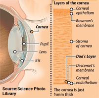
 The cornea is in front of the lens. It is a clear layer, seeming to lack substance. However, it is comprised of multiple layers and groups of cells and proteins which are highly organized.
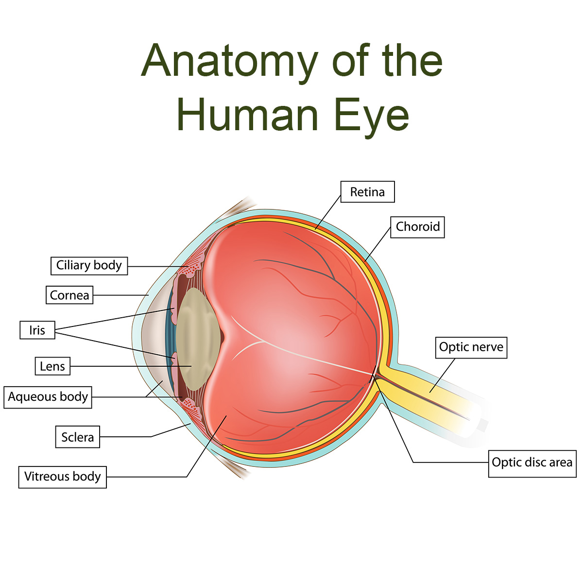
 Inflammation is a factor in many eye diseases, and what you eat affects inflammation. Whenever major holidays comes along, we are tempted to eat special foods. Many of these treats are high in calories and sugar, which contribute to inflammation. While you don’t want to spoil anyone’s fun, you can choose to eat foods that are anti-inflammatory. At the end of this article, you will find tips for limiting the damage while still enjoying the holiday.
  Have you ever heard that your eyes need plenty of nutrition? It’s true, and research backs this up. But did you know that the Standard American Diet tends to be very low in some eye-essential nutrients? The eyes are the second most physiologically active part of our body (#1 is the brain). At Natural Eye Care, we believe the eyes require approximately 25% of the nutrients we take into our body, if we eat a healthy diet. Even though most Americans consume enough calories, we may still have poor nutrition for the eyes. No wonder Macular Degeneration, cataracts, glaucoma, optic nerve
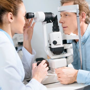
  Cornea transplant surgery involves removing a damaged or diseased cornea, and replacing it with tissue from a deceased donor. All or part of the cornea may be replaced. In the United States, eye surgeons do approximately 33,000 corneal transplants (keratoplasty) per year. The surgery might be recommended if vision is seriously impaired by scarring from an injury, or eye diseases such as Fuchs’ Dystrophy, Lattice Dystrophy, or Keratoconus.
 Keratoconus is a disorder of the cornea that causes visual distortion. Changes in the cellular structures of the cornea cause it to thin and bend into a pronounced cone shape, losing its normal gentle curvature. This leads to several types of visual distortion including blurring, halos around lights, and, in some cases, rapid vision loss. The signature sign of Keratoconus is the perception of multiple ghostly images, called monocular polyopia.